Hymenolepis nana- Dwarf Tapeworm
•
12 likes•4,166 views

Hymenolepiasis is caused by Hymenolepis nana (the dwarf tapeworm,a type of intestinal worm or helminth infecting humans, especially children.

Recommended
More Related Content
What's hot
What's hot (20)
Similar to Hymenolepis nana- Dwarf Tapeworm
Similar to Hymenolepis nana- Dwarf Tapeworm (20)
More from Anup Bajracharya
More from Anup Bajracharya (20)
Recently uploaded
Recently uploaded (20)
Hymenolepis nana- Dwarf Tapeworm
- 1. Hymenolepis nana Anup Muni Bajracharya
- 2. Causative agent • Hymenolepis nana • is also known as the dwarf tapeworm. • The species name ‘nana’ is derived from its small size which means dwarf of the adult worm. • It is the smallest and only cestode that parasites the human without any intermediate host. A.B
- 3. Habitat • In human: The adult worm resides in the ileal portion of the small intestine • It is also present in other mammals like rats and mice A.B
- 4. Geographical Distribution • The parasite is cosmopolitan in distribution. • The worm is found in temperate than tropical countries. • It is highly prevalent in • South Africa, • South Europe , • Latin America and • Middle-east Asia. A.B
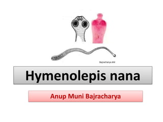
- 5. Morphology • small and thread like worms • measuring 1-4 cm in length with a diameter of 1mm. • Body consists of a scolex, a long neck and strobili consists of nearly 200 proglolttids. A.B
- 6. • Scolex: • globular, measuring 0.32 mm in diameter. • four suckers and a short rostellum armed with 20-30 spines in one ring. • Rostellum is retractable and always remains invaginated at the apex of the organ. • Neck: • It is relatively long and situated posterior to the head. A.B
- 7. • Strobila • consists of nearly 200 segments or proglottids. • Each mature segment- 0.3 mm in length by 0.9 mm breadth. • Proglottid contains both male and female reproductive organs. • Genital pores are marginal and are situated on the same side. • The uterus is a transverse sac with lobulated walls and there are three testes. • Proglottids neat the neck regions are small, short, narrow and immature and those away from the scolex are mature and gravid. • Segments at distal end are the gravid proglottids, which contain fertilized eggs. A.B
- 8. • Eggs • the infective form of parasite to human. • liberated in the faeces by gradual disintegration of the terminal segments. • colorless, spherical or oval in shape, • measuring 30-45mm in diameter. • has 2 distinct membranes. • Outer membrane -thin and colorless • On inner membrane there are two small “knobs” or poles from which 4– 8 filaments arise • Inner membrane (embryophore)- encloses an oncosphere with 3 pairs of lancet shaped hooklets that is • they are embryonated and have a 6- hooked oncospheres inside the shells. A.B
- 9. Life cycle • Life cycle is completed in a single host, man and does not require any intermediate host. • Man and rat act as both definitive and intermediate hosts. • Man acquires infections by injection of fully embroynated egg. • In the small intestine a hexacanth embryo is liberated. • It borrows into the villi of the anterior part of the small intestine and in about 4 days time develop into a typical larval stage called ‘cysticercoid’. • After reaching maturity the ileum ruptures and the larva enters the lumen of the small intestine. Later, it attaches to another villus further down and in the course of fortnight develops into adult worms. A.B
- 10. • The gravid proglolttids breaks off the adult worm and disintegrate to release embryonated eggs while it passes down the intestinal tract. • The eggs passed in the faeces are the source of infection for new host and the cycle is continued. • Sometimes, few eggs hatch out in the lumen of the small intestine and liberate embryos which directly invade the intestinal villi. • This phase of infection is called internal autoinfection and is responsible for increased worms load in intestine. • Some person infect own self by faeco-oral route due to bad personal hygiene. • This is called external manifestation and is responsible for persistence of the infection in a host. A.B
- 11. A.B
- 12. Mode of transmission • Infected human faeces are the chief source of infection. • Transmission of the man occurs- • Through faecel-oral route by ingestion of eggs from contaminated hands. • Frequently by contaminated food and water • Rarely from ingestion of food contaminated fleas habouring the cysticercoid larva. A.B
- 13. Pathogenesis • Even large numbers of parasite is well tolerated. • The mechanisms by which symptoms are usually produced is an allergic reaction. • In heavy infections large number of worms may cause: • Mechanical irritation of the intestine and produce various clinical manifestation and • Various allergic manifestation such as anal and nasal pruritus (redness ,itching) by releasing toxic metabolites. A.B
- 14. Clinical manifestation • Most infections are asymptomatic. • In heavy infections, symptoms include irritability, diarrhea, abdominal pain, sleep disorder, anal pruritus and nasal pruritus. • Rare symptoms anorexia, nausea and vomiting. • Complications • These include bloody diarrhea and behavioural disturbances. A.B
- 15. Laboratory diagnosis • 1.Specimen: • Stool sample • rectal swab • 2. Microscopy: • Diagnosis is based on the demonstration of characteristics eggs in the faeces by direct microscopy. • The eggs can readily be concentrated by the salt flotation and formalin ether sedimentation technique. • 3. Hematology: • Eosiniphila of more than 5% is seen in 1/3rd of the infected children • 4. Endoscopy: • Adult worm can be identified during endoscopic examination • 5. ELISA: • It has 80% sensitivity A.B
- 16. Treatment • Praziquantel: drug of choice • Albendazole, Mebendazole Prevention and control • Sanitary improvements • Uncontaminated food supplies • Rodent control in house • Personal hygiene A.B
- 17. THANK YOU ANY QUERIES ??? A.B