
Meek Micro-Skin Grafting and Acellular Dermal Matrix in Pediatric Patients: A Novel Approach to Massive Extravasation Injury
 , , ,
, , ,  ,
,
Abstract
:1. Introduction
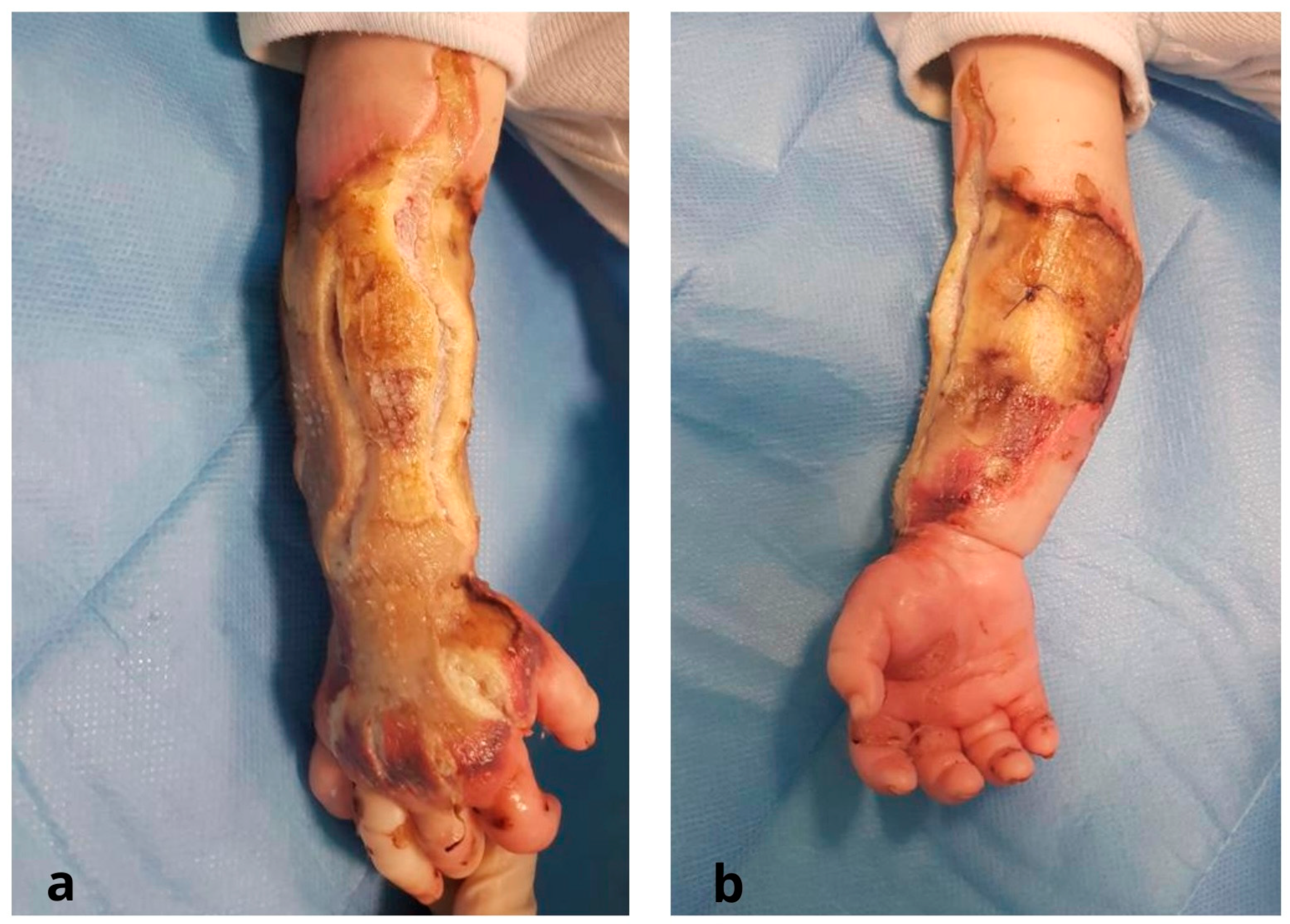
2. Case Report
3. Results
4. Discussion
5. Conclusions
Author Contributions
Funding
Institutional Review Board Statement
Informed Consent Statement
Data Availability Statement
Conflicts of Interest
References
- Hackenberg, R.K.; Kabir, K.; Müller, A.; Heydweiller, A.; Burger, C.; Welle, K. Extravasation Injuries of the Limbs in Neonates and Children—Development of a Treatment Algorithm. Dtsch. Arztebl. Int. 2021, 118, 547–554. [Google Scholar] [CrossRef] [PubMed]
- Wilkins, C.E.; Emmerson, A.J.B. Extravasation injuries on regional neonatal units. Arch. Dis. Child.-Fetal Neonatal Ed. 2004, 89, F274–F2755. [Google Scholar] [CrossRef] [PubMed] [Green Version]
- Falcone, P.A.; Barrall, D.T.; Jeyarajah, D.R.; Grossman, J.A. Nonoperative management of full-thickness intravenous extravasation injuries in premature neonates using enzymatic debridement. Ann. Plast. Surg. 1989, 22, 146–149. [Google Scholar] [CrossRef] [PubMed]
- Maruccia, M.; Ruggieri, M.; Onesti, M.G. Facial skin breakdown in patients with non-invasive ventilation devices: Report of two cases and indications for treatment and prevention. Int. Wound J. 2015, 12, 451–455. [Google Scholar] [CrossRef]
- Kostogloudis, N.; Demiri, E.; Tsimponis, A.; Dionyssiou, D.; Ioannidis, S.; Chatziioannidis, I.; Nikolaidis, N. Severe Extravasation Injuries in Neonates: A Report of 34 Cases. Pediatr. Dermatol. 2015, 32, 830–835. [Google Scholar] [CrossRef]
- Gault, D.T. Extravasation injuries. Br. J. Plast. Surg. 1993, 46, 91–96. [Google Scholar] [CrossRef]
- Hannon, M.G.; Lee, S.K. Extravasation injuries. J. Hand Surg. 2011, 36, 2060–2065. [Google Scholar] [CrossRef]
- Goverman, J.; Kraft, C.T.; Fagan, S.; Levi, B. Back Grafting the Split-Thickness Skin Graft Donor Site. J. Burn. Care Res. 2017, 38, e443–e449. [Google Scholar] [CrossRef] [Green Version]
- Rode, H.; Martinez, R.; Potgieter, D.; Adams, S.; Rogers, A.D. Experience and outcomes of micrografting for major paediatric burns. Burns 2017, 43, 1103–1110. [Google Scholar] [CrossRef]
- Maruccia, M.; Onesti, M.G.; Sorvillo, V.; Albano, A.; Dessy, L.A.; Carlesimo, B.; Tarallo, M.; Marcasciano, M.; Giudice, G.; Cigna, E.; et al. An Alternative Treatment Strategy for Complicated Chronic Wounds: Negative Pressure Therapy over Mesh Skin Graft. BioMed Res. Int. 2017, 2017, 8395219. [Google Scholar] [CrossRef]
- Medina, A.; Riegel, T.; Nystad, D.; Tredget, E.E. Modified Meek Micrografting Technique for Wound Coverage in Extensive Burn Injuries. J. Burn. Care Res. 2016, 37, 305–313. [Google Scholar] [CrossRef] [PubMed]
- Gacto-Sanchez, P. Surgical treatment and management of the severely burn patient: Review and update. Med. Intensiv. 2017, 41, 356–364. [Google Scholar] [CrossRef] [PubMed]
- Rijpma, D.; Claes, K.; Hoeksema, H.; de Decker, I.; Verbelen, J.; Monstrey, S.; Pijpe, A.; van Zuijlen, P.; Vries, A.M.-D. The Meek micrograft technique for burns; review on its outcomes: Searching for the superior skin grafting technique. Burns 2022, 48, 1287–1300. [Google Scholar] [CrossRef]
- Lee, S.Z.; Halim, A.S.; Wan Sulaiman, W.A.; Mat Saad, A.Z. Outcome of the Modified Meek Technique in the Management of Major Pediatric Burns. Ann. Plast. Surg. 2018, 81, 295–301. [Google Scholar] [CrossRef] [PubMed]
- Gagnier, J.J.; Kienle, G.; Altman, D.G.; Moher, D.; Sox, H.; Riley, D.; Allaire, A.; The CARE Group. The CARE guidelines: Consensus-based clinical case reporting guideline development. BMJ Case Rep. 2013, 2013, bcr2013201554. [Google Scholar] [CrossRef] [PubMed] [Green Version]
- Gierek, M.; Łabuś, W.; Słaboń, A.; Ziółkowska, K.; Ochała-Gierek, G.; Kitala, D.; Szyluk, K.; Niemiec, P. Co-Graft of Acellular Dermal Matrix and Split Thickness Skin Graft-A New Reconstructive Surgical Method in the Treatment of Hidradenitis Suppurativa. Bioengineering 2022, 9, 389. [Google Scholar] [CrossRef] [PubMed]
- Gierek, M.; Łabuś, W.; Kitala, D.; Lorek, A.; Ochała-Gierek, G.; Zagórska, K.M.; Waniczek, D.; Szyluk, K.; Niemiec, P. Human Acellular Dermal Matrix in Reconstructive Surgery-A Review. Biomedicines 2022, 10, 2870. [Google Scholar] [CrossRef]
- Cigna, E.; Maruccia, M.; Sorvillo, V.; Parisi, P.; Palumbo, F.; Onesti, M.G. The use of negative pressure therapy and hyaluronic acid for the management of post-traumatic lower limb injury. Int. Wound J. 2012, 10, 534–538. [Google Scholar] [CrossRef]
- Asuku, M.; Yu, T.-C.; Yan, Q.; Böing, E.; Hahn, H.; Hovland, S.; Donelan, M.B. Split-thickness skin graft donor-site morbidity: A systematic literature review. Burns 2021, 47, 1525–1546. [Google Scholar] [CrossRef]
- Kadam, D. Novel expansion techniques for skin grafts. Indian J. Plast. Surg. 2016, 49, 5–15. [Google Scholar] [CrossRef] [Green Version]
- Pope, E.R. Mesh skin grafting. Veter-Clin. N. Am. Small Anim. Pract. 1990, 20, 177–187. [Google Scholar] [CrossRef] [PubMed]
- Dahmardehei, M.; Vaghardoost, R.; Saboury, M.; Zarei, H.; Saboury, S.; Molaei, M.; Seyyedi, J.; Maleknejad, A.; Hospital, I.F. Comparison of Modified Meek Technique with Standard Mesh Method in Patients with Third Degree Burns. World J. Plast. Surg. 2020, 9, 267–273. [Google Scholar] [CrossRef] [PubMed]
- Menon, S.; Li, Z.; Harvey, J.G.; Holland, A.J.A. The use of the Meek technique in conjunction with cultured epithelial autograft in the management of major paediatric burns. Burns 2013, 39, 674–679. [Google Scholar] [CrossRef]
- Noureldin, M.A.; Said, T.A.; Makeen, K.; Kadry, H.M. Comparative study between skin micrografting (Meek technique) and meshed skin grafts in paediatric burns. Burns 2022, 48, 1632–1644. [Google Scholar] [CrossRef] [PubMed]
- Gierek, M.; Kawecki, M.; Mikuś, K.; Klama-Baryła, A.; Nowak, M. Biological dressings as a substitutes of the skin in the treatment of burn wounds. Ann. Surg. 2013, 85, 354–359. [Google Scholar] [CrossRef] [Green Version]
- Chong, S.J.; Choke, A.; Tan, B.K. Technical tips to enhance micrografting results in burn surgery. Burns 2017, 43, 983–986. [Google Scholar] [CrossRef]
- Kreis, R.W.; Mackie, D.P.; Vloemans, A.W.; Hermans, R.P.; Hoekstra, M.J. Widely expanded postage stamp skin grafts using a modified Meek technique in combination with an allograft overlay. Burns 1993, 19, 142–145. [Google Scholar] [CrossRef]
- Nicoletti, G.; Brenta, F.; Bleve, M.; Pellegatta, T.; Malovini, A.; Faga, A.; Perugini, P. Long-term in vivo assessment of bioengineered skin substitutes: A clinical study. J. Tissue Eng. Regen. Med. 2015, 9, 460–468. [Google Scholar] [CrossRef]
- Onesti, M.G.; Fioramonti, P.; Carella, S.; Maruccia, M. The importance of periwound skin in the treatment of “difficult wound”. G Chir. 2011, 32, 83–88. [Google Scholar]


| Diagnosis | Age (Weeks) | Tretment | Range of Motion |
|---|---|---|---|
| Inflamed Meckel diverticulum | 8 | Total parenteral nutrition (TPN) via intravenous access in the right forearm for maintenance purposes (8 weeks) | |
| Extensive Extravasation Injury of soft tissues and skin of the right forearm | 8 | Fasciotomies of both dorsum of the hand and forearm | |
| 8 | Multidisciplinary evaluation in Neonatal Intensive Care Unit (NICU) | ||
| 8 | Increased level of procalcitonin, PCR, D-Dimer, WBC, and body temperature. blood and urinary culture tests, rectal, pharyngeal, and auricular swabs for pathogen identification. Treatment with Amoxicillin and clavulanic acid (8–9 weeks) | ||
| 9 | Surgical debridement and temporary coverage with acellular dermal matrix (Pelnac®) | ||
| Extensive Extravasation Injury with Exposed Tendons and Underlying Tissues | 9 | Culture biopsies confirming enterobacter cloacae complex infection, leading to antibiotic regimen switch to Meropenem | |
| 9–12 | Dressings applied using vaseline gauzes and sterile gauze soaked in chlorhexidine | ||
| Appropriate Wound Bed Preparation and Coverage of Exposed Tendons and underlying tissues | 12 | Meek micrografting technique for split-thickness skin graft (STSG) expansion, using the right thigh as the donor site | |
| 13 | Engraftment of Meek micrografts | ||
| 13–15 | Re-epithelialization of each Meek graft occurred from its edges, gradually covering the wound bed. | ||
| Impaired Wrist and Elbow Joints, and Inability to Extend First Finger Impaired Wrist and Elbow Joints, and Inability to Extend First Finger | 15–41 | Follow-up with a comprehensive physical therapy program | Elbow Joint:
|
| 41–67 | Follow-up with a comprehensive physical therapy program | Elbow Joint:
|
Disclaimer/Publisher’s Note: The statements, opinions and data contained in all publications are solely those of the individual author(s) and contributor(s) and not of MDPI and/or the editor(s). MDPI and/or the editor(s) disclaim responsibility for any injury to people or property resulting from any ideas, methods, instructions or products referred to in the content. |
© 2023 by the authors. Licensee MDPI, Basel, Switzerland. This article is an open access article distributed under the terms and conditions of the Creative Commons Attribution (CC BY) license (https://creativecommons.org/licenses/by/4.0/).
Share and Cite
Maruccia, M.; Tedeschi, P.; Corrao, C.; Elia, R.; La Padula, S.; Di Summa, P.G.; Maggio, G.M.M.; Giudice, G. Meek Micro-Skin Grafting and Acellular Dermal Matrix in Pediatric Patients: A Novel Approach to Massive Extravasation Injury. J. Clin. Med. 2023, 12, 4587. https://doi.org/10.3390/jcm12144587
Maruccia M, Tedeschi P, Corrao C, Elia R, La Padula S, Di Summa PG, Maggio GMM, Giudice G. Meek Micro-Skin Grafting and Acellular Dermal Matrix in Pediatric Patients: A Novel Approach to Massive Extravasation Injury. Journal of Clinical Medicine. 2023; 12(14):4587. https://doi.org/10.3390/jcm12144587
Chicago/Turabian StyleMaruccia, Michele, Pasquale Tedeschi, Claudia Corrao, Rossella Elia, Simone La Padula, Pietro G. Di Summa, Giulio M. M. Maggio, and Giuseppe Giudice. 2023. "Meek Micro-Skin Grafting and Acellular Dermal Matrix in Pediatric Patients: A Novel Approach to Massive Extravasation Injury" Journal of Clinical Medicine 12, no. 14: 4587. https://doi.org/10.3390/jcm12144587