7-Figure Settlement: Animated Appendix Rupture
March 28, 2017
Appendicitis is a medical condition involving the inflammation of the appendix, a tube of tissue that extends from the large intestines. Surgery is required to remove it because if left untreated, the inflamed appendix will eventually burst or perforate, discharging infectious materials into the abdominal cavity.
A hospital and physicians admitted a patient with acute appendicitis and prepped him for urgent surgery. But instead of removing the entire appendix during the appendectomy operation, the hospital surgeon left a substantial portion of the appendix, which went on to rupture, leaking fecal matter into the patient’s abdomen for weeks.
The plaintiff started feeling fevers, chills, and a general feeling of profound discomfort. His wife brought him back to the Emergency Department 12 days later when his symptoms worsened. A CT scan showed three large collections of infectious fluid with air bubbles around his liver, cecum, and pelvis.
The patient was admitted into surgery for an exploratory laparotomy. Upon entering the patient’s abdomen, a foul stench filled the operating room. Surgeons reported seeing giant pools of fecal fluid, murky old blood, discharged pus, and a necrotic colon comprised of dead tissue that would need to be removed.
Stuart Ratzan, Esq., and Stuart Weissman, Esq., needed visuals that would show the egregious level of negligence involved in the first surgery, illustrate the disaster it created in the patient’s abdomen, and demonstrate the surgery needed to clean up the mess. We delivered an animation that would achieve the following visual objectives.
- Introduce viewers to the abdominal anatomy, and show what the patient's initial appendicitis looked like.
- Compare the correct way to remove it versus the incomplete appendectomy performed by the defendants.
- Illustrate how the appendix ruptured, filling the abdomen with infectious fecal matter.
- Compare the animated scene to a colorized CT scan taken by medical providers before the second surgery, confirming the locations of infectious matter.
- Break down the surgery that was required to suction out the murky fluid, remove a dead section of the patient’s colon, and reconnect the patient’s colon tube to restore intestinal continuity.
The following animation helped The Ratzan Law Group return an undisclosed, seven-figure settlement for the plaintiff.
COMPLETE ANIMATION
Watch the full animation above, and then check out its strategic breakdown below.
"While we resolved this case prior to trial, we used the animation to assist with negotiations. The defense knew we were serious about this case, and we were more than ready to gear up for trial. A huge thank you to the entire High Impact team."
Stuart Weissman, Esq., - The Ratzan Law Group
PART 1: INTRODUCING APPENDICITIS
The story begins with what the first surgery was supposed to address: the patient's inflamed appendix.

Normal Appendix VS Appendicitis
Comparing a normal appendix versus appendicitis helps establish why the initial surgery was needed in the first place.
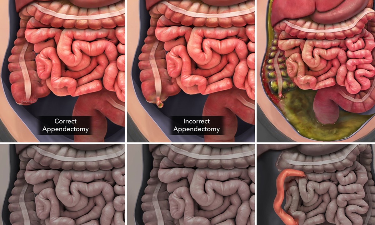
PART 2: COMPARING RIGHT VERSUS WRONG
After demonstrating why surgeons operated on the appendix, the next part shows the source of damages after they botched the surgery.

Correct VS Incorrect Appendectomy
Comparing the right way surgeons should have completed this surgery versus the way they neglected to complete it helped emphasize their clear negligence.
PART 3: ANIMATED APPENDIX RUPTURE
After establishing malpractice, we animated the progression of damages as the appendix rupture leaked fecal matter into the plaintiff's abdomen over the course of almost two weeks.

Animated Appendix Rupture Leaks Fecal Fluid for Two Weeks
Emphasizing the progression of damages helped capture the disturbing experience the plaintiff lived with for almost two weeks, as he was reporting fevers, chills, and a general feeling of profound discomfort.
PART 4: COLORIZED CONFIRMATION OF DAMAGE
After animating an illustrated depiction of overall damage, we display the actual CT scan taken in the ER alongside our artwork.

Comparing Illustrated Damage to Colorized CT-Scan
The colorized CT scan helped confirm the credibility of the art by depicting the same area of damage being portrayed in the illustration. Yellow was used in the CT scan to highlight areas of fecal matter and familiarize viewers with the otherwise black-and-white radiography.
PART 5: ANIMATED SURGERY
After both liability and physical damages had been established, we animated the surgical procedures required to clean out the mess, remove the plaintiff's dead colon tissue, and redirect the colon to restore intestinal continuity.

Animated Suction of Infectious Matter
First, surgeons needed to suck out all the infectious material that permeated throughout the plaintiff's abdominal anatomy.

Animated Resection of Dead Colon Tissue
The next part of the surgery involved the removal of the plaintiff's dead colon tissue, which posed an infectious risk to his anatomy.

Animated Anastomosis to Restore Intestinal Continuity
After removing part of the patient's colon, an anastomosis was needed to redirect his digestive system and restore intestinal continuity.

Animated Placement of Blake Drain
Finally, the animation concluded with the placement of a Blake Drain, which was needed to drain the area of any more infectious fluid and pus from the anatomy. Animation helps reinforce the reality of that situation.
High Impact’s team of visual strategists, artists and developers can build and customize your digital presentation for any case involving personal injury, medical malpractice, birth trauma - or any subject involving complex information. Contact us here or call (800) 749 2184 to learn more.