
Introduction
The posteromedial caecal wall, just below the end of the ileum, gives rise to the vermiform appendix, a small vermiform (worm-like) structure.1 The most frequent factor leading to emergency laparotomies has been identified as its inflammation. The primary role of the vermiform appendix, which also performs other gastrointestinal tract-related tasks, is immunological. Vermiform appendix's tonsil-like behaviour has also been claimed. Similar to how the tonsils protect the upper digestive system from pathogens, the vermiform appendix protects the small intestine from bacteria found in the large intestine.2 An organ with changeable location is the vermiform appendix. The location of the vermiform appendix is not governed by any set rules, however it is believed that it is strongly tied to the caecum's development.3 Recognizing the vermiform appendix variation; scientists have been studying its position as early as 1933. 4 The clinical manifestation of appendicitis is notoriously unpredictable due to variations in the location of the appendix, the patient's age, and the level of inflammation.5 Determining the appendix's typical position is crucial since it might create symptoms and signs specific to its position during appendicitis, which can cause it to resemble other conditions.6 We discovered that there is a dearth of material on this significant structure in terms of age while reading the literature. In light of this context, the current work is an effort to investigate the location and morphometry of the vermiform appendix in cadavers available in the anatomy department.
Aim
The aim of present study is to investigate certain morphological features of vermiform appendix in cadavers.
Objectives
To measure the different parameters of appendix such as length, diameter and distance of appendicular orifice from ileum.
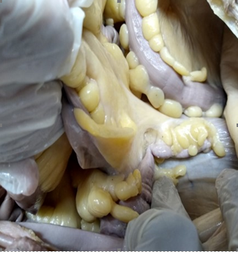
To note the different positions of appendix in cadavers.
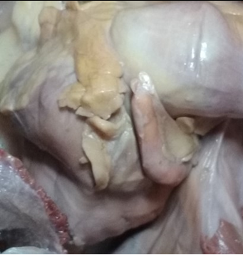
To see the variations of the mesoappendix.
To correlate the significance of these findings with clinical finding
Material and Methods Study
Sample size
50 CADAVERS available in the department of Anatomy will be utilized for study purpose.
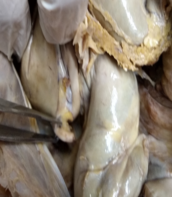
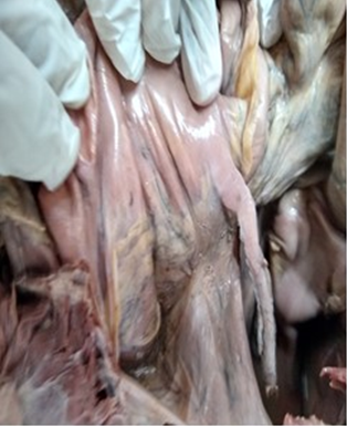
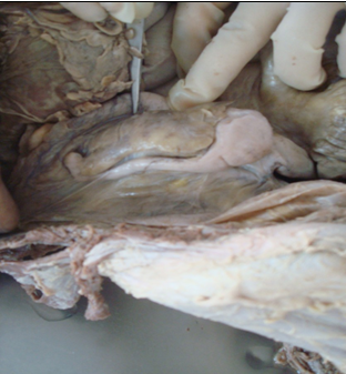
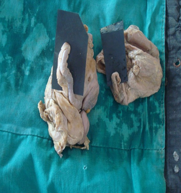
In each specimen the appendix was exposed and its position, length and diameter and the extent of mesoappendix was noted.
How to measure the vermiform appendix's length, Using a measuring tape with a centimetre graduation, the length of the vermiform appendix was determined by measuring the distance from the base to the tip of the appendix. Where the taeniae coli on the ascending colon and caecum merge is where the base of the appendix was found to be. The appendix serves as a point of reference for the anterior caecal taenia coli, which is typically identifiable.7 The findings of present work will be correlated with clinical findings in the light of available literature.
Results
Table 1
Different parameters of appendix
It is evident from table no I Length of appendix was 5.47± 1.59, diameter at base was 6.85±1.82, diameter at tip was 3.94± 1.11, distance of appendicular orifice below ileum was 1.14±0.85 and Distance of appendicular orifice below ileum inside was 2.19 ±0.78.
Table 2
Different positions of appendix
|
Positions |
Number |
Percentage |
|
Retrocaecal |
20 |
40% |
|
Pelvic |
15 |
30% |
|
Paracolic |
4 |
8% |
|
Splenic ( postileal ) |
4 |
8% |
|
Promonteric |
4 |
8% |
|
Midinguinal |
3 |
6% |
It is evident from this table that the most common position was retrocaecal 20(40%) followed by pelvic 15 (30%) least common was midinguinal.
Table 3
Different positions of Mesoappendix
|
Positions of Mesoappendix |
Number |
Percentage |
|
Mesoappendix reaches the tip |
37 |
74% |
|
Mesoappendix does not reach the tip |
13 |
26% |
It is evident from present study the most common position of mesoappendix was mesoappendix reaches the tip 37 (74%) followed by mesoappendix does not reaching the tip was 13 (26%)

Discussion
Most people think of the vermiform appendix as a vestigial organ, and the only reason it's important in surgery is because of its predisposition for inflammation, which causes the clinical illness known as acute appendicitis. The most frequent cause of "acute abdomen" in young teenagers is acute appendicitis, and an appendectomy is frequently the first major procedure carried out by a surgeon-in-training. The clinical presentation of appendicitis is notoriously unpredictable due to variations in the position of the appendix, the patient's age, and the level of inflammation.8, 9, 10 Misdiagnosis rates range from 10% to 33% depending on the age group.11 The diagnosis of appendicitis remains primarily clinical and requires a combination of observations, clinical judgement, and surgical sense despite tremendous breakthroughs in current radiography imaging and diagnostic laboratory studies.
Paul et al studied Sixty (60) human post mortem vermiform appendix, age ranging from 0 to 65 years. Length of vermiform appendix decreases gradually with increasing age and was highly significant7 (P<0.001) when compared between the groups.12
Golalipour, M. J et al observed The average length of appendix was 6.61cms in males and 6.06 cms in females. Pelvic position was the predominant position (in 33.3%) followed by retrocaecal in 32.4%, preileal in 18.8% and subcaecal in 12.8% respectively. In 65.8% of cases, the mesoappendix failed to reach appendicular tip. Retrocaecal and pelvic positions were the most common position in Turkman and native Fars races, respectively.13
Arindom et al carried out study on 25 cadavers where he found length of appendix was more in male cadavers than female.14 The length of appendix was 6.3 ±2.08 SD close to our study. In present study the length of appendix was 5.47± 1.59 (Table 1) which is close to Golalipour, M. J et al.13 Syed et al.15 found adult male length of appendix was 7.72 cm and adult female length of appendix it was 6.93 cm which is similar to our study.
In present study diameter at base was 6.85±1.82, diameter at tip was 3.94± 1.11, distance of appendicular orifice below ileum was 1.14±0.85 and Distance of appendicular orifice below ileum inside was 2.19 ±0.78 these measurements could not be compared with other studies as we did not come across any reference with these measurements.
In present study the most common position was retrocaecal 20(40%) followed by pelvic 15 (30%) least common was midinguinal which was similar to Arindom et al 14, 15 Retrocecal position was common in Syed et al study and least common was preileal and postileal. Retrocaecal was similar to our study. We have not observed postileal position in our study. Ehab I. El-Amin et al16 appendix was found in the retrocaecal position (60%), which is the normal developmental position of the appendix, followed by pelvis position (35.0%), and the pre-ileal position (1.7%).
In present study the most common position of mesoappendix was mesoappendix reaches the tip 37 (74%) followed by mesoappendix does not reaching the tip was 13 (26%). Arindom et al 14 found complete in four cases and in 21 cases it is incomplete which is reverse of our cases. Syed et al observed in 17 cases it reached the tip of appendix and in 33 cases it failed to reach the tip of appendix.
Conclusion
This study provide gross anatomical information for the surgeons and to contribute to the medical world for updating the data. Having standard data on the vermiform appendix is useful for clinicians as well as anthropologists. The findings of the present study can provide information about morphologic variations of the appendix in western Indian population. However, further studies with a larger sample size are required to make better decision.


