Microscopy is a vital tool used in scientific research and analysis. It involves the use of specialized instruments to observe and study microscopic objects or structures. There are different types of microscopes available, but two of the most commonly used are light microscopy and electron microscopy. In this article, we will explore the differences between these two techniques and their applications.
|
Table of contents
|
Light Microscopy
 |
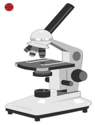
| Light Microscope |
Light microscopy, also known as optical microscopy, is a type of microscopy that uses visible light to illuminate and magnify specimens. It is the most widely used microscopy technique, and it is relatively easy to use and cost-effective compared to other types of microscopy.
Light microscopy can be divided into two main categories: bright-field microscopy and fluorescence microscopy. In bright-field microscopy, light passes through the specimen and is then refracted or absorbed, creating contrast that allows the specimen to be seen. Fluorescence microscopy, on the other hand, uses a fluorescent dye that emits light when exposed to a particular wavelength of light, which allows specific structures or molecules to be labeled and visualized.
The major advantage of light microscopy is that it can be used to observe living cells and organisms without damaging them. It is also useful for observing a wide range of structures and is an essential tool in fields such as cell biology, microbiology, and histology.
Electron Microscopy
 |
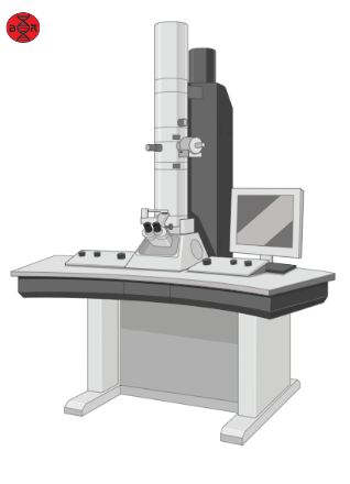
| Transmission Electron Microscope (TEM) |
Electron microscopy, on the other hand, uses a beam of electrons instead of light to illuminate specimens. This technique can achieve much higher magnifications and resolutions than light microscopy, making it possible to visualize smaller structures and details in much greater detail.
There are two main types of electron microscopy: transmission electron microscopy (TEM) and scanning electron microscopy (SEM). In TEM, electrons are transmitted through a thin specimen, and the resulting image is formed by detecting the electrons that pass through the specimen. In SEM, a beam of electrons is scanned across the surface of a specimen, and the resulting image is formed by detecting the electrons that are scattered back from the specimen’s surface.
One of the main advantages of electron microscopy is that it can visualize structures at a much higher magnification and resolution than light microscopy, allowing for a more detailed understanding of the microscopic world. However, electron microscopy requires specialized equipment and techniques, and it can be expensive and time-consuming to use. Additionally, electron microscopy typically requires specimens to be fixed and prepared in a way that can destroy the natural structure of the specimen, making it unsuitable for observing living cells and organisms.
Applications
Both light microscopy and electron microscopy have different applications and are used in different scientific fields. Light microscopy is ideal for studying living cells and organisms and is widely used in fields such as cell biology, microbiology, and histology. Electron microscopy, on the other hand, is best suited for studying non-living specimens and is commonly used in fields such as material science, nanotechnology, and structural biology.
In conclusion, both light microscopy and electron microscopy are valuable tools in scientific research and analysis. Light microscopy is useful for observing living cells and organisms and is easy to use and cost-effective. Electron microscopy, on the other hand, can achieve much higher magnifications and resolutions than light microscopy and is ideal for studying non-living specimens. By understanding the differences between these techniques, scientists can choose the right tool for their research and achieve a more detailed understanding of the microscopic world.
Source: ChatGPT as a part of our AI Articles feature.
Images: Created with BioRender.com
One thought on “Light Microscopy Vs Electron Microscopy – Understanding the Differences and Applications”
Comments are closed.