Approach Considerations
No specific laboratory tests confirm or exclude the diagnosis of thromboangiitis obliterans (TAO; also known as Buerger disease).
Arteriographic abnormalities consistent with TAO are sometimes seen in limbs that are not yet clinically involved; therefore, arteriography of all four limbs may be required. Echocardiography and computed tomography (CT) angiography (CTA) should always be performed in patients thought to have TAO in order to exclude a proximal source of thromboemboli or atheroemboli as the cause of distal vessel occlusion.
Laboratory Studies
The primary goal of a laboratory workup in patients thought to have the disease is to exclude other disease processes in the differential diagnosis. Tests often used as markers for the diagnosis of systemic vasculitis, such as the acute-phase reactants, yield negative results in patients with TAO. Tests commonly ordered include the following:
-
Complete blood count (CBC) with differential
-
Liver function tests
-
Renal function tests
-
Urinalysis
-
Glucose (fasting)
-
Erythrocyte sedimentation rate (ESR)
-
C-reactive protein (CRP)
-
Antinuclear antibody (ANA)
-
Rheumatoid factor (RF)
-
Complement
-
Anticentromere antibody
-
Scl-70 antibody
-
Antiphospholipid antibodies
Arteriography
The hallmark angiographic findings in patients with TAO are nonatherosclerotic, segmental occlusive lesions of the small and medium-sized vessels (eg, digital, palmar, plantar, tibial, peroneal, radial, and ulnar arteries) with formation of distinctive small collateral vessels around areas of occlusion, known as corkscrew collaterals (see the image below). These corkscrew collaterals represent dilated vasa vasorum of the occluded arteries, which are now serving to provide distal perfusion, albeit at significantly higher resistance than the native vasculature.
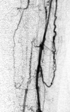 Lower-extremity arteriogram of peroneal and tibial arteries of patient with thromboangiitis obliterans (Buerger disease) demonstrates classic findings of multiple small and medium-sized arterial occlusions with formation of compensatory "corkscrew collaterals."
Such arteriographic findings suggest TAO but are not pathognomonic, because similar lesions can be observed in patients with scleroderma, CREST (calcinosis cutis, Raynaud phenomenon, esophageal motility disorder, sclerodactyly, and telangiectasia) syndrome, systemic lupus erythematosus, rheumatoid vasculitis, mixed connective-tissue disease, antiphospholipid-antibody syndrome, and even diabetes mellitus.
Histologic Findings
In its acute phase, TAO is characterized by highly cellular, segmental, occlusive, inflammatory thrombi, with minimal inflammation in the walls of affected blood vessels. Secondary spread from the affected small and medium-sized arteries to contiguous veins and nerves is often observed. Microscopically, the polymorphonuclear leukocyte (PMN)-predominant inflammatory cellular aggregate may form microabscesses and multinucleated giant cells.
In the subacute phase, intraluminal thrombosis progressively organizes, but it may defer to vascular recanalization. [10] The end-stage phase of TAO is characterized by mature thrombus and vascular fibrosis.
In all three stages of the disease, the integrity of the normal structure of the vessel wall, including the internal elastic lamina, is maintained. This maintenance of structural integrity distinguishes TAO from arteriosclerosis and from other types of systemic vasculitis, in which disruption of the internal elastic lamina and the media can be extensive.
-
Feet of patient with thromboangiitis obliterans (Buerger disease). Note ischemic ulcers on distal portion of left great, second, and fifth toes. Although patient's right foot is normal in gross appearance, angiography demonstrated compromised arterial flow to both feet.
-
Superficial thrombophlebitis of great toe in patient with thromboangiitis obliterans (Buerger disease).
-
Tobacco smoke stains on male patient's fingers suggest diagnosis of thromboangiitis obliterans (Buerger disease). Patient presented with small, painful ulcers on tips of thumb and ring finger.
-
Lower-extremity arteriogram of peroneal and tibial arteries of patient with thromboangiitis obliterans (Buerger disease) demonstrates classic findings of multiple small and medium-sized arterial occlusions with formation of compensatory "corkscrew collaterals."
Tables
Positive Criterion |
Positive Points |
||
Age at onset |
< 30 y (+2) 30-40 y (+1) |
||
Foot intermittent claudication |
Present (+2) By history only (+1) |
||
Upper extremity |
Symptomatic (+2) Asymptomatic (+1) |
||
Migrating superficial thrombophlebitis |
Present (+2) By history only (+1) |
||
Raynaud phenomenon |
Present (+2) By history only (+1) |
||
Angiography; biopsy |
If typical, both (+2) Either(+1) |
||
Negative Criterion |
Negative Points |
||
Age at onset |
45-50 y (−1) >50 y (−2) |
||
Sex; smoking |
Female (−1) Nonsmoker (−2) |
||
Location |
Single limb (−1) No lower extremity involved (−2) |
||
Absent pulses |
Brachial (−1) Femoral (−2) |
||
Arteriosclerosis, diabetes, hypertension, hyperlipidemia |
Discovered 5.1-10 y after diagnosis (−1) Discovered 2.1-5 y later (−2) |
||
Score |
Probability of Diagnosis |
0-1 |
Diagnosis excluded |
2-3 |
Diagnosis suspected (low probability) |
4-5 |
Diagnosis probable (medium probability) |
≥6 |
Diagnosis definite (high probability) |


